X-ray computed microtomography (GTK)
X-ray computed microtomography (μ-CT) is a non-destructive technology that precisely images the interior of solid materials, e.g.s rocks in 3D at any orientation. The device installed at GTK is a GE phoenix v|tome|x s with a 240 kV direct microfocus tube (smallest voxel size 5 μm) and a 180 kV transmission nanofocus tube (smallest voxel size 900 nm). A large 2024x2024 pixel detector enables fast scan times and scanning of large samples, up to 30 cm in diameter and 41 cm in length, weighing up to 10 kg. The connected computer systems are up to the demanding tasks of data processing and visualization, using the new PerGeos software from FEI, which was developed from the top of the line visualization software, Avizo.
The GTK device also has a Deben CT5000TEC sample stage (in maintenance as of July 19th 2018) and metrology|ct, helix|ct and offset|ct extensions, which allow accurate voxel size calibration, helical scanning to continuously scan long samples and eliminate Feldkamp artefcats, and offset scanning to effectively enlarge the detector width to 3800 pixels and thus allow a better FOV to resolution ratio or a larger sample diameter, respectively.
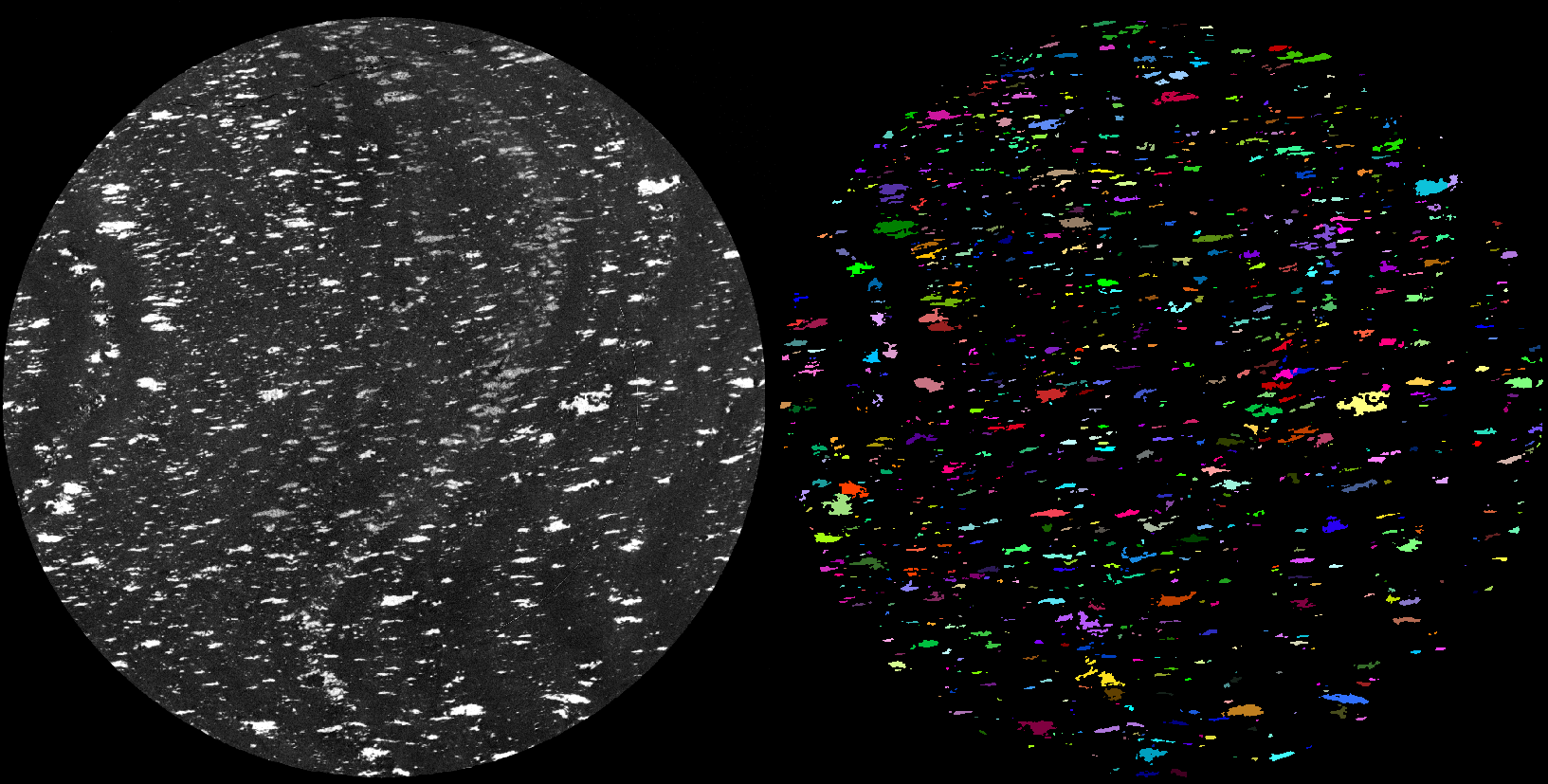
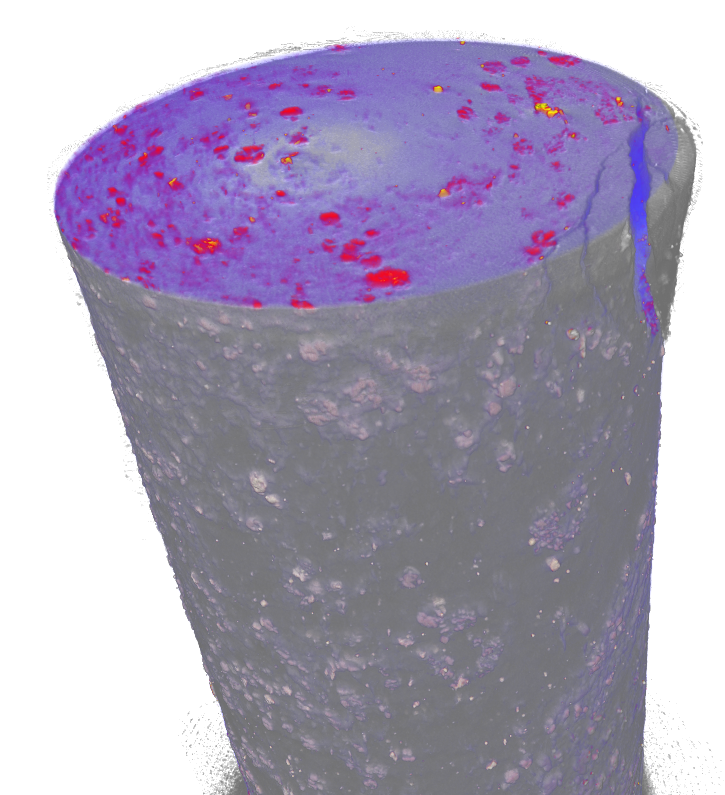
Figure 1. Left panel: Tomographic slice from an oriented drill core with high density grains segmented and separated for analysis. Right panel: Drill core sample scanned before (red and blue) and after (grayscale) it fractured near the top.
In the modern geoscience research, this equipment is essential, e.g. in the 3D visualization and analysis of gold distribution in a rock sample, Platinum-Group Minerals and other precious metal-bearing minerals with common non-metallic mineral phases, rock textures, micro-sedimentary features, glacial sediments, glaciolacustrine varves, brittle-ductile fractures in rocks and crystals, porosity and permeability analysis, sizes, shapes and orietations of crystals/grains, morphologies of fossils, soils and clays, metallurgy and metal products, building materials, and industrial waste.
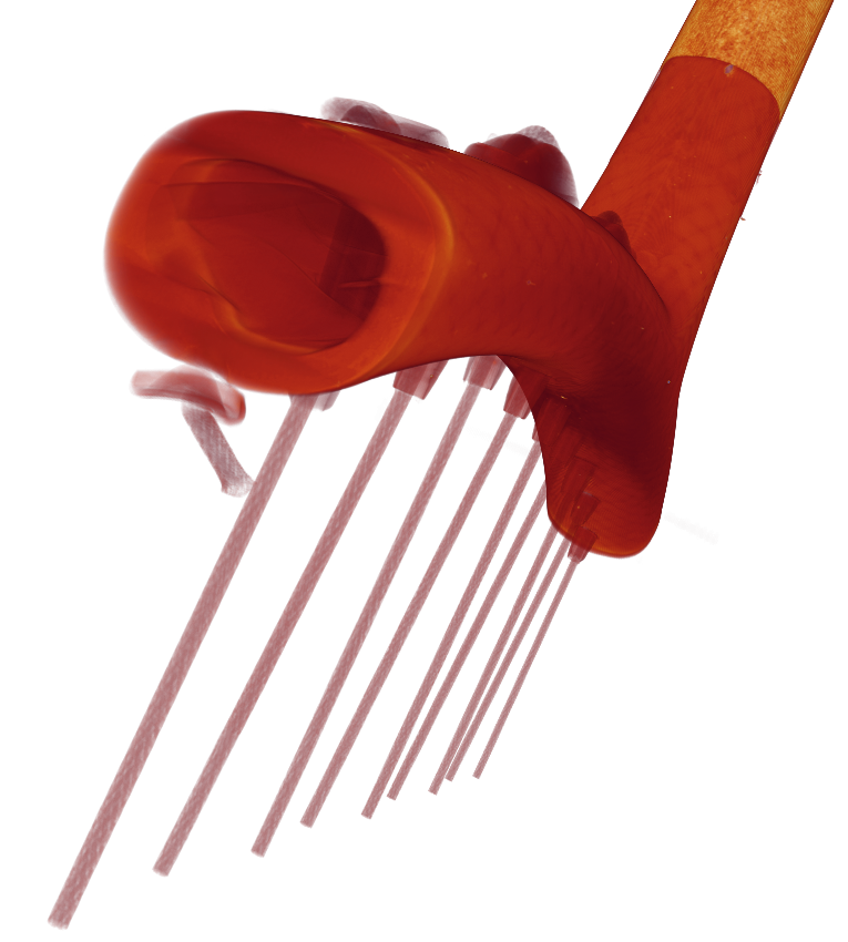
Figure 2. The joint between the head and shaft of a badminton racket.
In addition, the method is highly versatile and especially important in applied geosciences and in rock mechanics and engineering, the oil industry, archaeology, biological sciences and in electronics. With the appropriate acceleration voltage this method can reveal internal details down to a sub-micrometer resolution of a given sample. The device perfectly complements other tomography devices in Finland. List of other domestic devices, along with more information on tomography on a national scale, can be found on the website of FinTomo (in Finnish), a national tomography network where GTK is a founding member.
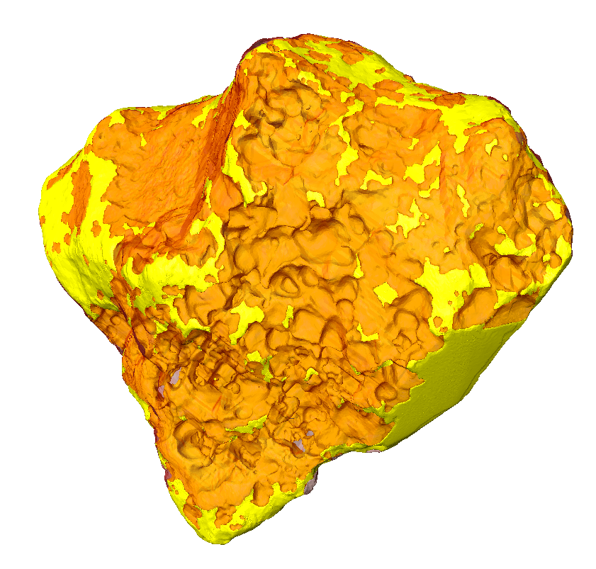
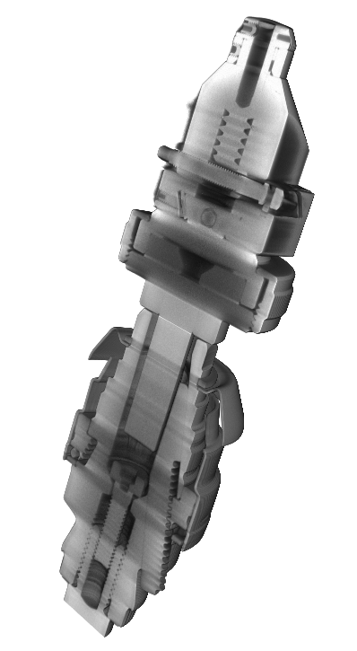
Figure 3. Left panel: Iron meteorite, showing the silicate (orange) and Fe-Ni (yellow) phases. Right panel: Drill chuck and a gonyometer, showing the inner workings of both.
As of September 20th, 2019, the GTK device has logged 780 hours on 509 scans on the microfocus tube and 198 hours on 47 scans on the nanofocus tube. There have been 69 users from 26 institutions in 9 countries. Users from all institutions are welcome and are encouraged to contact the lab manager Jukka Kuva (firstname.lastname@gtk.fi).
Pricing, list of lab publications, etc., can be found on the XCT lab website.


